
Oral cancer screening
By Alderwood Family Dentistry • March 23, 2019
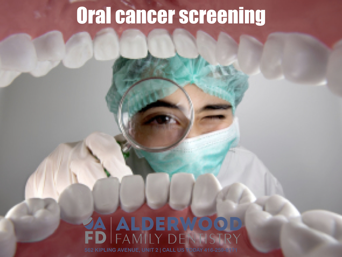
What to expect during an oral cancer screening
Oral cancer screening is quick and easy, with no pain or discomfort involved. It’s critical for early diagnosis and prevention, and it takes less than five minutes, also, it can be done by your dental hygienist or dentist at your regular check-up appointment.
Approximately 3,200 new cases of oral cancer will be diagnosed in Canada this year and many more cases will be diagnosed with dysplasia (pre-cancer), according to the British Columbia Cancer Research Centre. Oral cancer is the sixth most common type of cancer in North America. Canadian statistics reveal that more cases of oral cancer are diagnosed in a year than cervical or ovarian cancer, and more people die from oral cancer in a year than from melanoma or cervical cancer.
The good news?
Approximately 84 percent of oral cancer cases may be detected early by a dental professional.
Your dental professional will be looking for any lumps or abnormalities, red, white or grey areas and/or tenderness.
What to expect
Your dental professional should:
- Physically examine and inspect the outside and inside of your gums and cheeks, extending all the way from top to bottom and front to back.
- Examine and inspect the floor of your mouth underneath your tongue and the roof of your mouth.
- Touch and inspect the lymph nodes in your neck and jaw, examine the inside and outside of your lips, your major salivary glands (cheeks and floor of mouth), as well as your jaw joint (TMJ) and the area around your ears.
- Visually inspect your face for signs of asymmetry or swelling.
- Pull out tongue with gauze and inspect posterior, sides, bottom and top.
- Have you extend your tongue and say “ahhh” in order to inspect the back of your mouth and your throat.
- Ask about your smoking and/or drinking habits, your lifestyle and any specific health conditions you have or medications you are taking which could affect the condition of your mouth or head and neck area.
Our office use a scope, called Velscope, with a special light that will show cancerous and precancerous lesions as dark areas.
Screenings should be done at every six month check-up.
If you feel that a screening has not been done for awhile, or if you have not gotten one at all, voice your concerns and ask for one. All dental hygienists and dentists are trained to do oral cancer screenings.
Performing an oral cancer self-check
If you only visit the dentist once a year or less, here’s how to perform a self-check, and what to watch for between screenings:
- First, arm yourself with one of those handy oral mirrors available at any drugstore. You’ll be able to inspect your mouth much more easily.
- Make sure you have a bright light. A small flashlight works well.
- Check for any new lumps, bumps or unusual tenderness anywhere in your mouth. You can check by palpating the tissue between your thumb and forefinger, or pressing against your face from the outside (similar to checking for breast lumps). For the floor of your mouth, press beneath your tongue.
- To check your tongue, grasp it with a gauze or cloth and extend it (the posterior is a common site for oral cancer). Say “ahhh” to check the back of your tongue and throat (you may need to ask for some help here).
- Visually check your whole mouth, watching for any discoloured areas, or spots with unusual texture. If you check regularly, you will soon be able to differentiate between what’s normal and what’s not.
- In general, follow as closely as possible the procedure carried out in the dental office.
- Be aware of any sore spots in your mouth or throat that don’t heal within two weeks, or if you have difficulty swallowing. Check with your dentist or doctor right away.
If a suspicious area is found, it does not mean you have oral cancer. Only a small number of people with abnormalities will be diagnosed with oral cancer. However, all abnormal areas need further investigation to rule out precancerous or cancerous lesions, and this may include a biopsy of the suspicious area.